Structure of a Skeletal Muscle Fiber. Students are directed to draw label and color code their model of a sarcomere as well as muscle contraction.
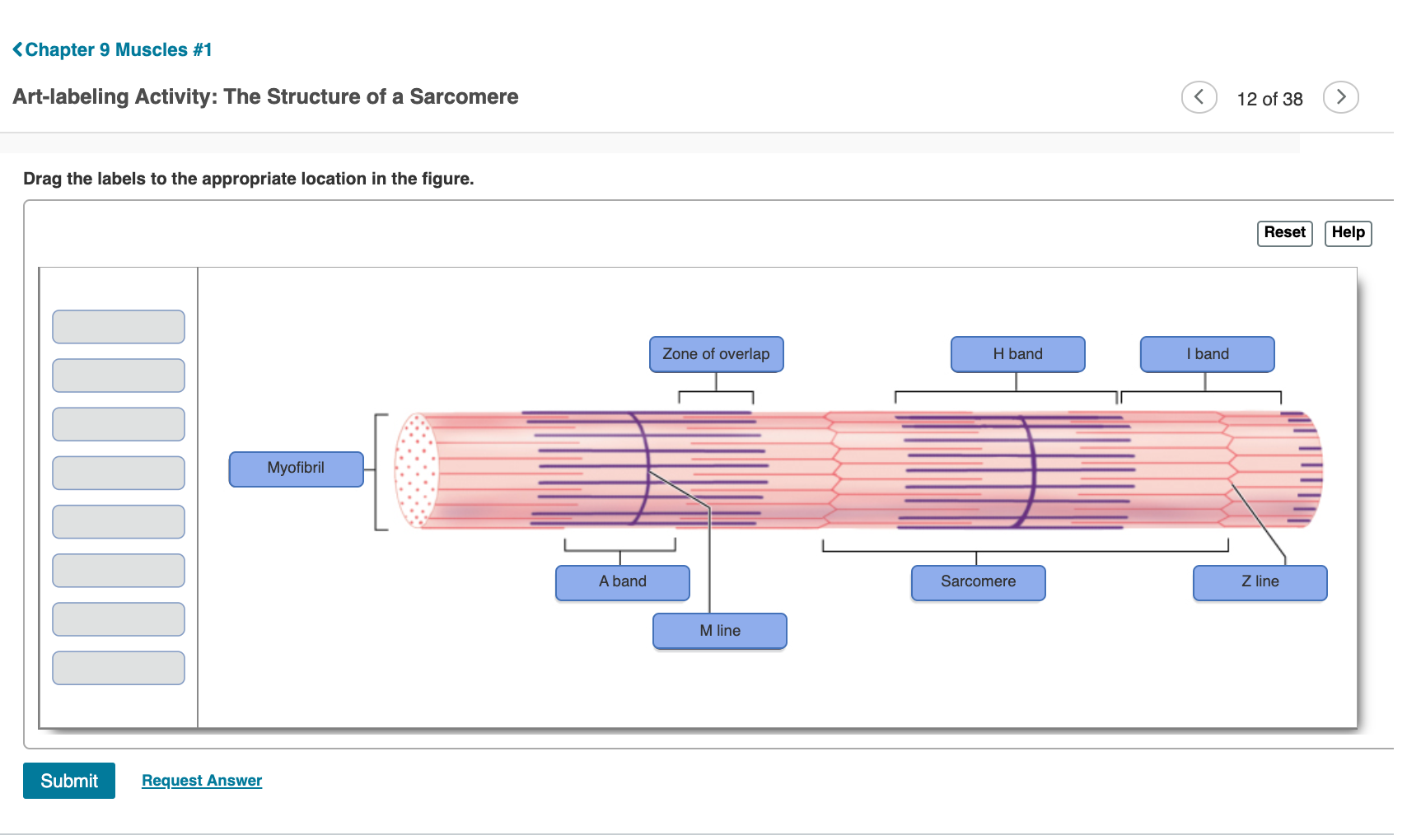
Solved Chapter 9 Muscles 1 Art Labeling Activity The Chegg Com
Start studying Art Labeling Activity.

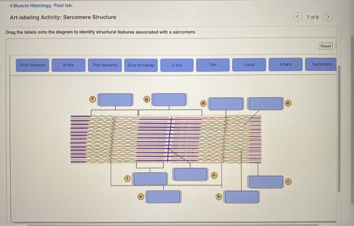
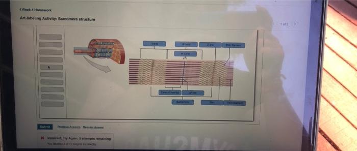
. Sarcomere Structure Drag the labels onto the diagram to identity structural features associated with a sarcomere. PPT - Chapter 47 Care of the Patient with a Blood or. The Spiral Organ of the Cochlea.
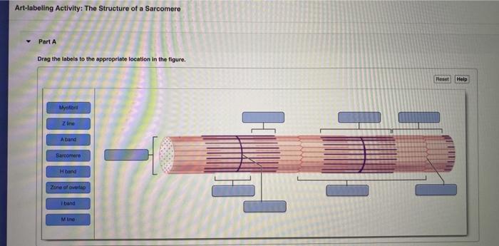
The Structure Of A Sarcomere Part A Drag The Labels To The Appropriate Location In The Figure. 42322 503 PM Week 3 Chapter 9 810 Correct Art-labeling Activity. Start studying Art-Labeling Activity.
Actin A Band Mline Z A. 41722 334 PM Ch 09 HW Ch 09 HW Due. When the sarcomere contracts and shortens__________.
Sarcomere structure Label the parts of a sarcomere. Reset Help A Band Barmere Hand Band MI Art-Labeling Activity. Learn vocabulary terms and more with flashcards games and other study tools.
Reset Help A band bend Thinaments Mline Thick framanta Sarcomero Zine Tion Zone of overlap O Subna Request Answer. The pinkish hue of individuals with fair skin is the result of the crimson color of oxygenated hemoglobin contained in red blood cells circulating in the dermal capillaries and reflecting through the epidermis. The Structure Of A Skeletal Muscle Fiber Part A Drag The Labels Onto The Diagram To Identity Structural Features Associated With A Skeletal Muscle Fiber.
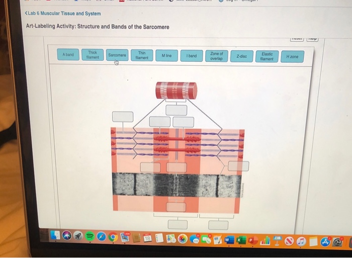
A sarcomere is defined as the region of a myofibril contained between two cytoskeletal structures called Z-discs also called Z-lines and the striated appearance of skeletal muscle fibers is due to the arrangement of the thick and thin myofilaments within each sarcomere Figure 1022. Help Reset Help Reset Applied force Resistance Fulcrum Second class Third class First class I band A band Z line Titin H band Zone of overlap M line Sarcomere Thin. Identify the structure of the muscle fiber as indicated.
Learn vocabulary terms and more with flashcards games and other study tools. Reset Help A Band Barmere Hand Band MI Art-Labeling Activity. This will allow students to get creative and master the muscle content.
A group of sarcomere comprises a myofibril within a muscle. A sarcomere is the basic functional unit of any muscle. 33 Label The Lymphatic System - Labels Design Ideas 2020.
Start studying Art-labeling Activity. The striated appearance of skeletal muscle fibers is due to the arrangement of the myofilaments of actin and myosin in sequential order from one end of the muscle fiber to the other. The thick filaments in the A band and thin filaments in the I band interact and.
Skeletal Muscle Structure And Contraction Bio103 Human Biology - Identify the structure of the muscle fiber as indicated. Lab - Integumentary System 326 Correct Art-Labeling Activity. The Structure Of A Sarcomere Part A Drag The Labels To The Appropriate Location In The Figure.
The sarcomere is the contraction unit in the skeletal muscles that are under the control of motor. The Structure Of A Sarcomere Part A Drag The Labels To The Appropriate Location In The Figure. Learn vocabulary terms and more with flashcards games and other study tools.
The Structure Of A Skeletal Muscle Fiber Part A Drag The Labels Onto. The Structure of a Skeletal Muscle Fiber. Art Labeling Activity.
Reset Help A Band Barmere Hand Band MI Art-Labeling Activity. NUK Simply Natural Glass Baby Bottle The NUK brand is designed to keep growing. Structure of the epidermis PartA Drag the appropriate labels to their.
A sarcomere is the area between two Z lines that can be regarded the fundamental structural and functional unit of muscle tissue. Label different areas of an individual muscle unit known as a sarcomere below. 35 Label The Parts Of The Lymphatic System - Labels Information List.
View Ch 09 HWpdf from BIO 231 at Springfield Technical Community College. 1159pm on Sunday April 3. The storage and release of calcium ions is the key function of the.
Review sarcomere structures and the components of muscle contraction with this informal assessmenthomeworkactivity. CISES 16-6 B On This Picture Draw And Label The F. Part A Drag the labels to the appropriate location in the figure.
The Structure Of A Skeletal Muscle Fiber Part A Drag The Labels Onto. The A band stays the same. The Anatomy of the Ear external and middle ear.
Each packet of these microfilaments and their regulatory proteins troponin and tropomyosin along with other proteins is called a sarcomere. 33 Label The Lymphatic System. A group of skeletal muscle fibers together with the surrounding perimysium form a n.
The Z-line establishes the sarcomeres lateral borders and anchors the thin titin and nebulin filaments. After learning about sarcomeres one of the most. The structure of.
Solved Art Labeling Activity The Structure Of A Sarcomere Chegg Com
Solved Art Labeling Activity Course Hero

Art Labeling Activity The Structure Of A Skeletal Muscle Fiber Diagram Quizlet

Art Labeling Activity Sarcomere Structure Diagram Quizlet
Solved Art Labeling Activity Sarcomere Structure Drag The Chegg Com
Solved Muscle Histology Post Lab Art Labeling Activity Chegg Com
Solved Lab 6 Muscular Tissue And System Art Labeling Chegg Com
Solved Week 4 Howwork Art Labeling Activity Sarcomere Chegg Com
0 comments
Post a Comment